American Journal of Ophthalmology AJO 2917 Page 9

9
HIGASHIDE, OCT and Angiography of Toxocara Granuloma
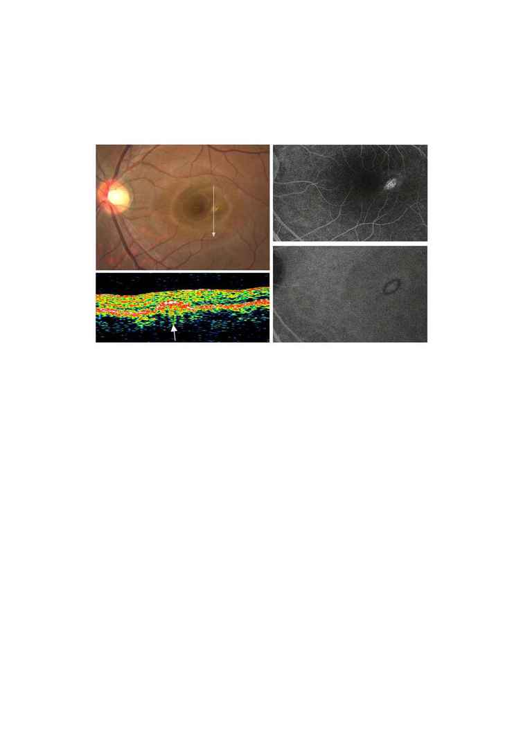
FIGURE 2. The Toxocara granuloma 3 months after onset.
(Top left) Fundus photograph shows a pigmented lesion without
exudation. The arrow indicates the location and direction of the scan
by OCT.
(Top right) Late phase fluorescein angiogram shows an oval-shaped
reticular hyperfluorescent lesion surrounded by a hypofluorescent
rim with minimal dye leakage.
