American Journal of Ophthalmology AJO 2917 Page 7

7
HIGASHIDE, OCT and Angiography of Toxocara Granuloma
Figure Legends
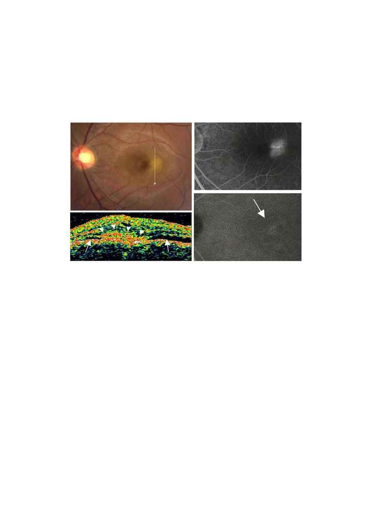
FIGURE 1. The Toxocara granuloma at the initial examination.
(Top left) Fundus photograph shows an ill-defined yellowish elevated
lesion with serous retinal detachment. The direction of the optical
coherence tomographic (OCT) scan is shown by the arrow.
(Top right) Late phase fluorescein angiogram shows dye leakage from
the center of the macular lesion.
(Bottom left) OCT shows a highly reflective mass protruding above
